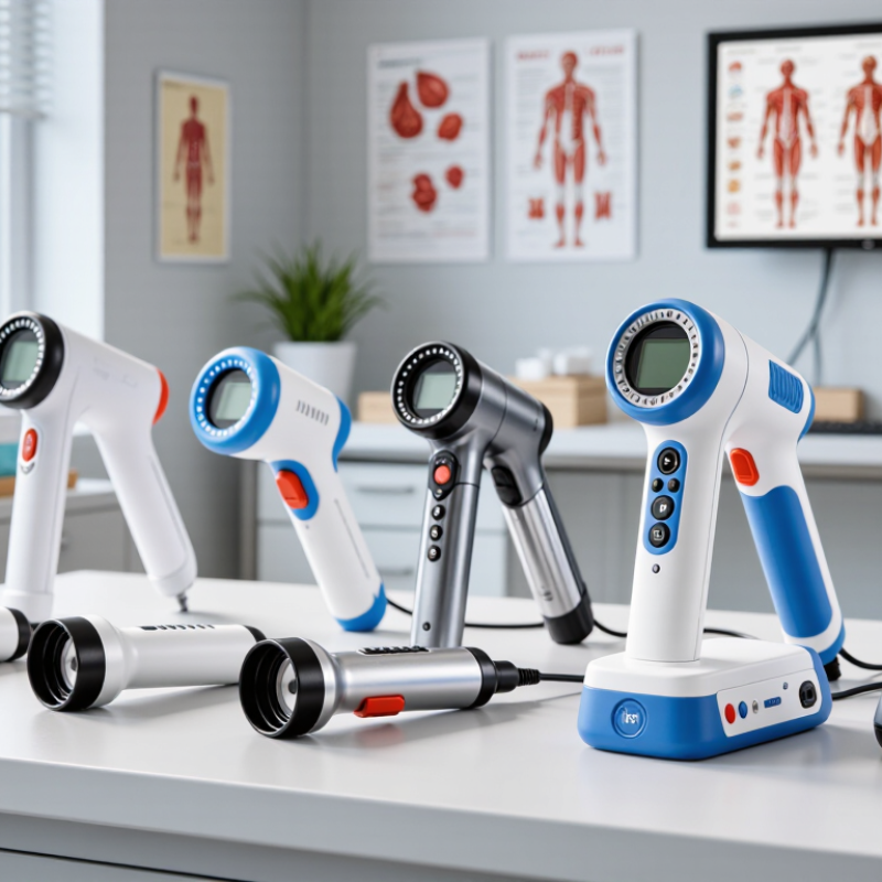
| 1 | Basic Portable Examination Model
★★★★★ | 3×–5× fixed or selectable magnification | Approximately 200–300 mm | White LED; adjustable intensity; coaxial or ring illumination | Integrated digital camera or external smartphone mount; still images and video | Approximately 0.5–1.0 kg | Rechargeable battery or USB power; compact carrying case | Primary-care screening, outreach clinics, and mobile examinations | Confirm that the light is centered on the examination field and that the device can be operated comfortably with one hand. |
| 2 | Variable-Magnification Diagnostic Model
★★★★★ | 5×, 10×, and 15× selectable magnification | Approximately 200–400 mm | High-intensity white LED with smooth brightness control | HD image capture, video recording, on-screen measurement, and file export | Approximately 0.8–1.5 kg | Rechargeable battery; AC adapter for extended sessions | Routine diagnostic assessment and follow-up documentation | Check whether magnification changes preserve focus and whether image files can be exported in standard formats such as JPEG or MP4. |
| 3 | Smartphone- or Tablet-Connected Model
★★★★☆ | 3×–10× optical magnification | Approximately 150–300 mm | Integrated white LED; brightness controlled through the device or handle | Wireless or wired connection to Android or iOS devices; image sharing and teleconsultation support | Approximately 0.4–0.9 kg, excluding the mobile device | Battery-powered; commonly charged through USB | Telemedicine, remote supervision, training, and distributed screening programs | Verify operating-system compatibility, data privacy controls, connection stability, and availability of offline image storage. |
| 4 | High-Resolution Video Documentation Model
★★★★☆ | 5×–15× optical or digital-assisted magnification | Approximately 250–400 mm | High-CRI white LED with adjustable intensity | Full HD or higher video output; snapshot button; external monitor or computer connection | Approximately 1.0–1.8 kg | AC and rechargeable-battery operation, depending on configuration | Clinical records, education, quality audits, and specialist review | Assess actual image sharpness, frame rate, glare control, and the time required to transfer or archive examination files. |
| 5 | Filter-Enabled Assessment Model
★★★★☆ | 5×–15×, with fixed or selectable levels | Approximately 200–350 mm | White LED with green or blue-green filter option | Digital still images and video; filter selection shown in the examination workflow | Approximately 0.8–1.6 kg | Rechargeable or mains-powered; suitable for fixed clinical rooms | Detailed visualization of vascular patterns and tissue features during clinical assessment | Confirm that the filter can be engaged without losing focus, reducing brightness excessively, or introducing color distortion. |
| 6 | Rugged Outreach and Field Model
★★★★☆ | 3×–10× magnification | Approximately 200–350 mm | Energy-efficient LED with protected optical housing | Basic image capture through an integrated camera or mobile-device adapter | Approximately 0.7–1.5 kg | Long-life rechargeable battery; protective case; optional vehicle or USB charging | Rural clinics, humanitarian programs, community screening, and locations with unstable power | Prioritize battery endurance, dust and moisture protection, shock resistance, spare-part availability, and ease of cleaning. |
| 7 | Lightweight Entry-Level Model
★★★☆☆ | 2×–5× magnification | Approximately 150–250 mm | Built-in white LED with manual brightness adjustment | Optional smartphone adapter or basic digital output | Approximately 0.3–0.8 kg | Battery-powered or USB rechargeable; minimal setup required | Training, low-volume clinics, preliminary examinations, and budget-sensitive procurement | Check optical clarity, illumination uniformity, battery replacement options, and whether accessories are included or sold separately. |